

How to Read Your Dental Report
Start with Dental X-rays: Understanding Wisdom Teeth, Cavities, Periodontitis, and Implant Follow-ups
After getting dental X-rays, many people look at a row of black-and-white teeth and still can't tell what the dentist is pointing at. In fact, when reading a report, you can start with three questions: which tooth, what was found, and what needs to be confirmed or done next.
This article is based on the video "An In-Depth Explanation of How to Read Dental X-rays" by Dr. Lin Junsheng, a dentist in Shenzhen. All images are actual screenshots from the original video, with timestamps in the captions. The video focuses on explaining X-rays; the report-reading steps and medical notes here are supplementary general information, not a diagnosis for any patient.
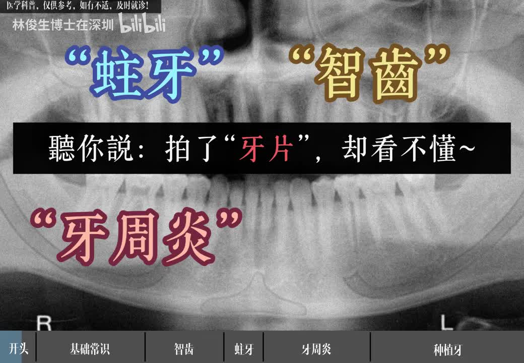
Figure 1 | Original video 00:06: Wisdom teeth, cavities, and periodontitis are common topics when reading dental X-rays.
1. First, Tell Apart the X-ray, the Imaging Report, and the Treatment Plan
A dental X-ray is an image; an imaging report describes the structures and abnormalities seen on it; the final diagnosis and treatment plan also depend on symptoms, an oral exam, and any other needed tests. You won't always get a separate written imaging report; sometimes the dentist explains the image directly.
You can read it in this order:
- Check the details. Confirm the name, date, and type of exam to make sure it's from this visit.
- Confirm the location. Ask the dentist to match the tooth numbers in the report to your mouth and the image. Left and right are from the patient's point of view; check the orientation markers on the image rather than guessing from the screen.
- Find the findings. Note whether the abnormality is in the crown, the root, the surrounding bone, or a gap where a tooth is missing.
- Read the tone of the conclusion. Words like "consider," "suspected," or "correlate clinically" mean more evaluation is needed; they don't equal a confirmed diagnosis.
- Clarify the next steps. What needs treatment, what can be watched, are more tests needed, and when is the follow-up?
Dental X-rays can help find problems that are hard to see in a visual exam, but whether you need them, and how often, should be decided by your dentist based on symptoms and risk. American Dental Association: Dental X-rays
2. What Do Black and White Mean on a Dental X-ray?
The video first explains the black-and-white image and tooth structure. Think of it this way: different tissues block X-rays to different degrees, so they appear as different shades of gray. Hard tooth tissue usually looks whiter, while areas like the pulp chamber look relatively darker.
"Black" doesn't mean a cavity, and "white" doesn't mean healthy. Normal structures naturally vary in brightness, and the image is also affected by technique, overlapping structures, and display settings. The video's line "the whiter, the harder" is fine as a first approximation, but you can't use it to measure tooth hardness.
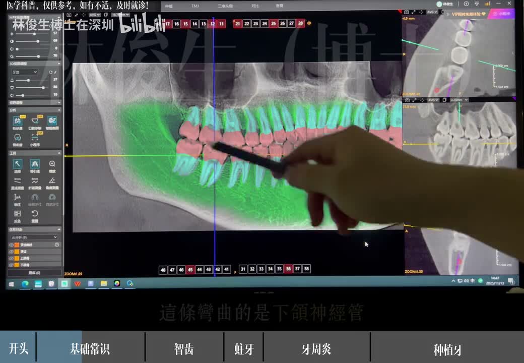
Figure 2 | Original video 00:34: Colored markings help explain the teeth and surrounding bone; these colors are annotations, not the color of any disease.
Three locations are enough to remember: the crown is the part visible in the mouth that does the chewing; the root is buried in the bone; the pulp chamber is the space inside the tooth that holds nerves, blood vessels, and other tissue. The video also marks the mandibular canal area with a yellow line, a reminder that when removing a lower wisdom tooth, its relationship to the roots matters.
3. Four Common Types of Dental X-rays, Each for Different Questions
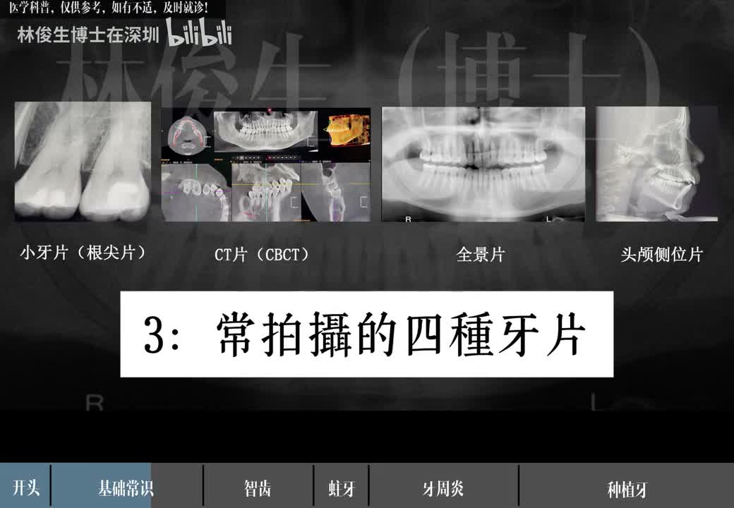
Figure 3 | Original video 00:46: Periapical X-ray, CBCT, panoramic X-ray, and lateral cephalometric X-ray.
| Exam type | Use in the video | What to ask when you get the report |
|---|---|---|
| Periapical X-ray, a.k.a. the "small film" | Looks at a single tooth or a small area | Is there anything abnormal at this tooth's root or around the root tip? |
| Panoramic X-ray | An overview of all teeth and the jaws | Where are the wisdom teeth? Any missing teeth or obvious bone changes? |
| CBCT (cone-beam CT) | Evaluates complex cases in 3D | Why is a 3D scan needed? What specific question will it answer? |
| Lateral cephalometric X-ray | Described in the video as commonly used for orthodontics | How does the relationship between my teeth and jaws affect the orthodontic plan? |
Choose the exam based on a specific question. CBCT provides 3D information but involves radiation exposure, so it should be used only when clinically needed, not routinely just to "see more." FDA: Dental CBCT
4. Wisdom Teeth: Check the Position First, Then Discuss Extraction
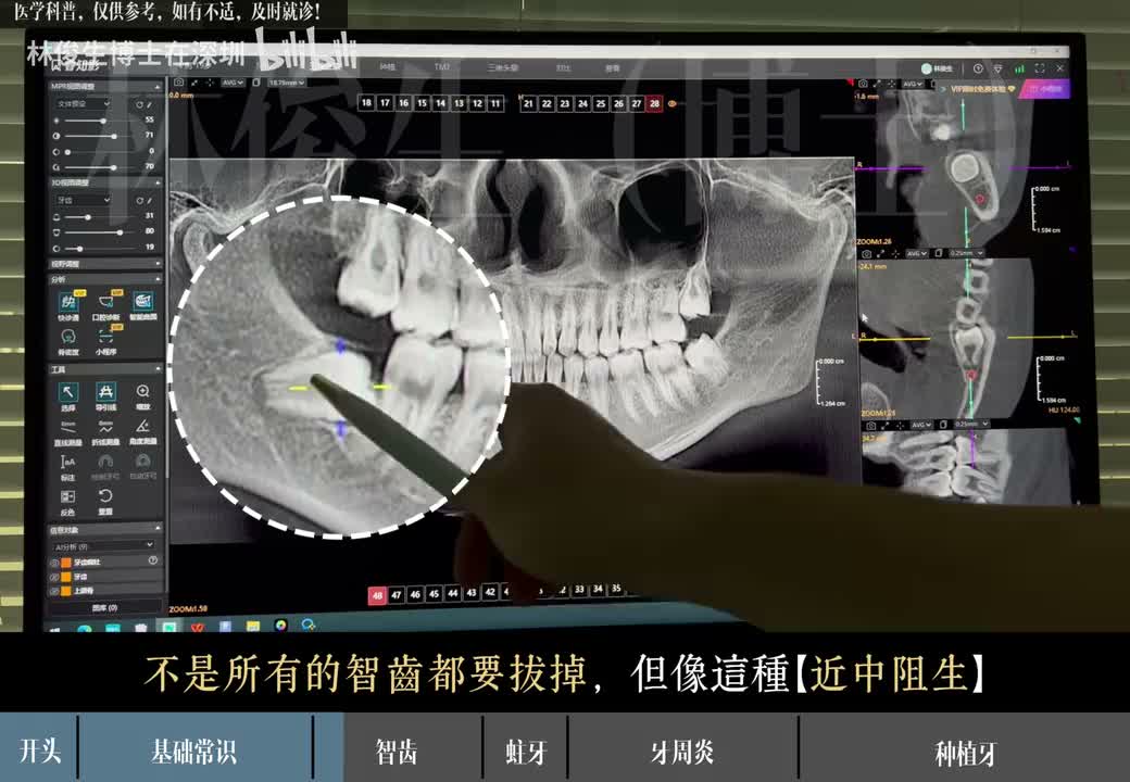
Figure 4 | Original video 01:10: The presenter zooms in on a wisdom tooth tilted toward the neighboring tooth in front, called "mesial impaction."
"Impacted" means a tooth hasn't erupted normally. The video uses a wisdom tooth tilted toward its neighbor to show that the area may be hard to clean, get inflamed repeatedly, or affect the neighboring tooth. It also shows a fairly upright wisdom tooth on the other side, stressing that not all wisdom teeth need to come out.
If your report says "impacted wisdom tooth," keep asking: Is it causing pain, infection, or problems with the neighboring tooth? How close are the roots to the nerve canal? What are the risks of keeping and watching it versus removing it? "It's growing crooked" alone can't decide extraction for everyone. NHS: Wisdom tooth removal
5. Cavities: Focus on How Deep the Problem Goes, Not Just Dark Spots
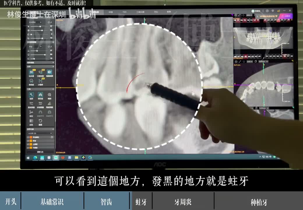
Figure 5 | Original video 01:42: The presenter points to a darker area on the crown and, in this case, explains it as a cavity.
The video explains decay using an image where part of the crown is darker and the structure looks broken. When reading a report, focus on: which tooth and which surface, how large the area is, and how close it is to the pulp chamber. A dark area on the image is only a clue; the dentist still needs to confirm it with an exam. Don't mistake the normal pulp chamber or overlapping structures for a cavity.
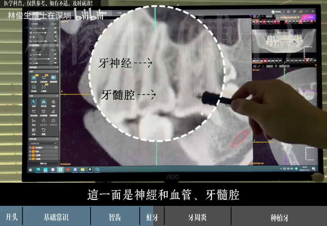
Figure 6 | Original video 01:50: The image labels the "dental nerve" and "pulp chamber" to show the space and tissue inside a tooth.
The video links deep cavities to root canal treatment. More precisely, whether you need a root canal depends on the state of the pulp and whether the tooth can be saved, not just on looking "close to the nerve" on an X-ray. Root canal treatment mainly deals with inflamed or infected pulp tissue inside the tooth. American Association of Endodontists: Root canal treatment
You can ask your dentist directly: "Can this be filled now, or does the pulp need further evaluation? What's the basis for that judgment?"
6. Periodontitis: Look at the Supporting Bone, and Check the Gums Too
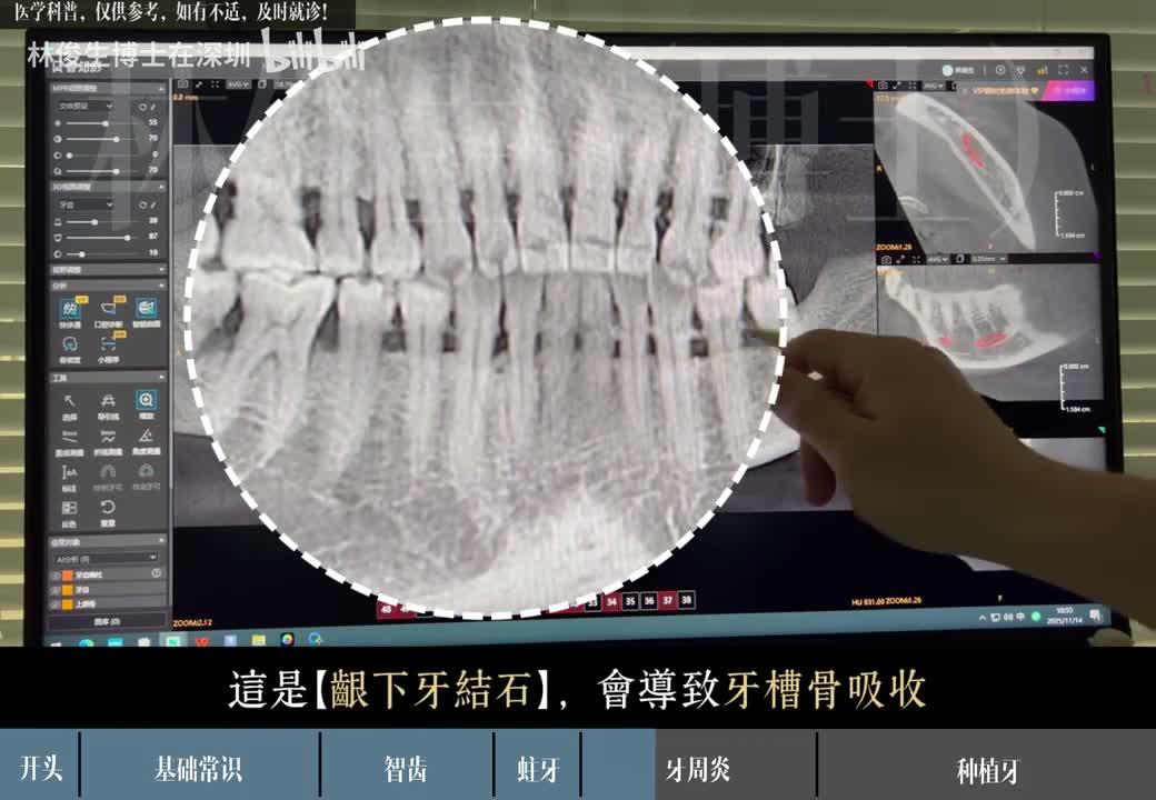
Figure 7 | Original video 02:14: The presenter interprets some projections near the tooth neck as subgingival tartar and discusses alveolar bone loss.
The video uses this case to explain how tartar, inflammation, alveolar bone loss, and loose teeth are related. If a report mentions "alveolar bone loss," it means there are signs that the bone supporting the teeth has been lost; but how severe it is and whether it's still progressing require a clinical exam and, where needed, comparison with earlier images.
A periodontal exam isn't just about X-rays; it also includes checking gum inflammation, bleeding, and probing periodontal pockets. X-rays help show bone loss but can't replace a periodontal exam. NIDCR: Diagnosing gum disease
The video also raises the question "why do the gaps between my teeth feel bigger after a cleaning?" If this happens, ask your dentist to explain the state of your gums and bone; it doesn't mean the cleaning damaged your teeth. The case in the video needed periodontal treatment, and the video introduces subgingival scaling; actual treatment and follow-up intervals should be based on your condition. NIDCR: Treating gum disease
7. Missing Teeth and Implants: X-rays Show More Than "Is There a Tooth"
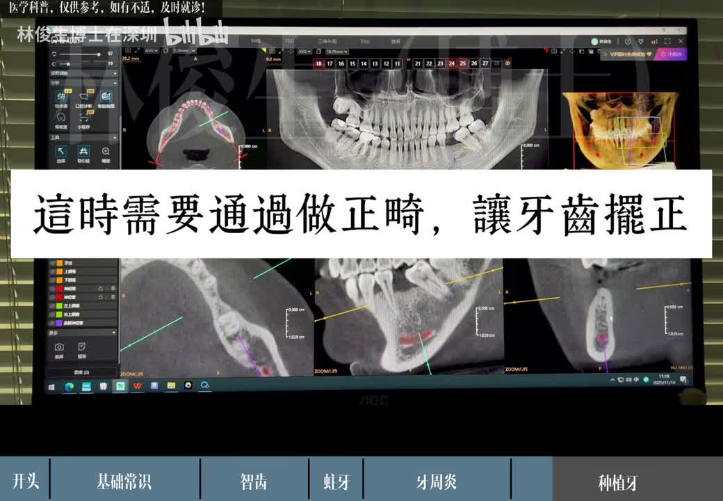
Figure 8 | Original video 03:02: Because the neighboring teeth had tilted and there wasn't enough space for a restoration, the presenter suggests first adjusting tooth positions with orthodontics.
The long-term missing-tooth case in the video involves tilted neighboring teeth, insufficient restoration space, and bone assessment. It reminds us that treating a gap requires considering space, neighboring teeth, bone, and the final restoration together. The orthodontics and bone grafting mentioned are this case's plan and shouldn't be copied as required steps for every patient with a missing tooth.
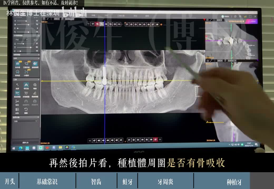
Figure 9 | Original video 03:38: The presenter explains checking for bone loss around the implant at a follow-up.
Finally, the video shows a follow-up about three years after an implant: asking whether food gets stuck easily, checking the bite, and looking at the bone around the implant. When reading a follow-up report, you can ask: "Has anything changed compared with before? Does this need to be judged together with an oral exam? Should my cleaning routine or follow-up plan change?"
The video mentions floss, water flossers, and regular check-ups. Ask your dentist to recommend specific tools, techniques, and follow-up frequency based on your restorations and oral health; there's no need to fix it at one X-ray per year on your own.
8. Bring These Five Questions to Your Follow-up Visit
- Which tooth is the report talking about? Please point to it on the X-ray and in my mouth.
- Is this a confirmed problem, or an imaging clue that needs further investigation?
- What other oral exams are needed to decide? What would additional imaging answer?
- What are the pros and cons of treatment, watching, or other options?
- When should I come back, and what changes should make me contact you sooner?
The point of understanding your report is to be able to discuss the problem clearly with your dentist. The images in this article are example cases from the video and can't be used to judge your own teeth; if you have any discomfort, see a dentist promptly.
Video and image source: Dr. Lin Junsheng (dentist in Shenzhen), "An In-Depth Explanation of How to Read Dental X-rays", about 3 minutes 59 seconds long. Screenshots keep the original frames and creator watermark, and caption timestamps match the video. This article is a topical rewrite, not a word-for-word transcript; links to authoritative sources are used to supplement and qualify the medical statements. Compiled and fact-checked on September 13, 2026.